
It’s important to note that not all clinics/hospitals have all of these types of MRI machines. Some facilities only have traditional closed MRIs. That being said, it’s always good to ask about your options, especially if you are claustrophobic, obese, or have difficulty laying down flat for a long period of time.
What is an MRI?
Magnetic resonance imaging (MRI) is a type of scan that uses strong magnetic fields and radio waves to produce detailed images of the inside of the body. MRI is a non-invasive imaging technology that produces three dimensional detailed anatomical images without the use of damaging radiation. An MRI scanner is a large tube that contains powerful magnets. You lie inside the tube during the scan. A patient has to lay very still during the imaging process in order not to blur the image.
An MRI generates a static electromagnetic field that realigns a small fraction of the hydrogen protons inside water molecules. Once those protons are lined up, coils in the scanner emit a short burst of radio-frequency waves that cause the protons’ magnetic fields to wobble. When the radio burst ends, the protons release energy, sending out a faint echo of the radio waves that is detected by receiver coils and gives a picture of the anatomy being scanned.
What is an MRI used for?
They work the best for non-bony parts or soft tissues of the body. The brain, spinal cord and nerves, as well as muscles, ligaments, and tendons are seen much more clearly with MRI than with regular x-rays or CT Scan. In the brain, MRI can differentiate between white matter, grey matter, and cerebral spinal fluid in structural images of the brain. It can also be used to diagnose aneurysms and tumors. An MRI is the imaging modality of choice when frequent imaging is required for diagnosis or therapy, especially in the brain.
What is the difference between an open MRI and a closed MRI?
A closed MRI is a machine that takes detailed images of your anatomy in a narrow cylindrical container. A closed MRI requires you to lie completely still.
An open MRI is open on the sides or features wider openings, still requiring the patient to lie on a sliding table. An open MRI was created to offer an alternative for patients who either have symptoms of anxiety, suffer from claustrophobia, or due to to them being obese and unable to fit in a closed MRI. While the open MRI does a great job at providing comfort, it does not provide the same level of detail as a closed MRI at this time. It produces less detailed images because the open nature of the machine does not provide as strong of a magnetic field. 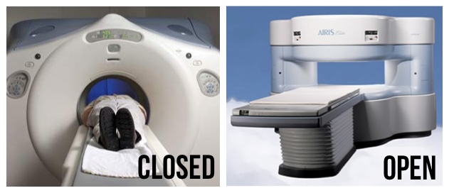
What is a 3 Tesla MRI?
This type of closed MRI uses magnetic fields that have double the strength of a traditional MRI machine, thus producing an even more detailed image in less time. The unit of measurement used to quantify the strength of a magnetic field in an MRI machine is called a Tesla (T). Most MRI scanners operate at a strength of 1.5 Tesla. A 3 Tesla MRI, however, operates at twice the normal strength, providing a greater signal-to-noise ratio, which is a major determinant in generating the highest quality image.
The higher resolution of the 3 Tesla MRI produces more detailed images, which are beneficial when diagnosing pathological conditions involving the brain, spine, and musculoskeletal system. The resolution and clarity also allow radiologists to identify smaller lesions and anatomical structures that cannot be seen with less powerful machines. This MRI allows for more sophisticated imaging procedures with more accurate diagnosis and lowers the risk of distorted images, thus eliminating the need for repeated scans.
What is an Extremity MRI?
This is a diagnostic imaging procedure that uses a closed MRI machine to look at the tissues in the arms and legs. Unlike a traditional MRI procedure that uses a large tube-shaped device, an extremity MRI uses a smaller scanner designed specifically for the body’s extremities. This eliminates the potential for claustrophobia, which some patients experience when enclosed in a full-body MRI machine. an extremity MRI also won’t limit your body movements quite as much as a closed MRI does. This type of scan is used specifically for diagnostic imaging of the arm, leg, hand, or foot. The machine uses radio waves and a magnetic field to generate images of the inside of the extremity in order to diagnose problems with the muscles, bones, joints, nerves, or blood vessels.


What is a Stand Up or Upright MRI?
Open Upright MRI scanners also offer the ability to capture images of your body in various positions, to diagnose the pain or discomfort you are experiencing. Depending on the source of our pain, you may be scanned sitting, standing, bending or twisting. Standing, bending, or stretching can cause pain for some patients, but the pain diminishes when lying down (during the scan). As a result, the cause of the pain may not be as visible in a recumbent scan, and therefore, not found
This MRI has the ability to perform weight bearing positional studies for the spine, shoulder, pelvis, SI joints, knee, ankle, feet, hips, and brain; the ability to perform studies in flexion, extension, rotation, and lateral bending positions; Upright MRI can evaluate effects of gravity on Pelvic Viscera for patients with suspected bladder, uterus, and rectal prolapsed – without preparation or probing – making the study truly comfortable for the patient; the ability to perform prostate studies for patients without preparation or probing, making the study truly comfortable for the patient; the upright MRI has higher field strength than any other “open” MRI scanners, which allows for higher resolution and enhanced image quality; this MRI also ideal for larger patients contingent on height and weight.


What is an fMRI?
One kind of specific and specialized MRI is functional Magnetic Resonance Imaging (fMRI.) This is used to observe brain structures and determine which areas of the brain consume more oxygen (activate) during various cognitive tasks. It is used to advance the understanding of brain organization and offers a tool for assessing neurological status and neurosurgical risks.
Functional magnetic resonance imaging (fMRI) measures the small changes in blood flow that occur with brain activity. fMRI may detect abnormalities within the brain that cannot be found with other imaging techniques.
Physicians perform fMRI to:
- examine the functional anatomy of the brain.
- determine which part of the brain is handling critical functions such as thought, speech, movement and sensation, which is called brain mapping.
- help assess the effects of stroke, trauma, or degenerative disease (such as Alzheimer’s) on brain function.
- monitor the growth and function of brain tumors.
- guide the planning of surgery, radiation therapy, or other invasive treatments for the brain.
Various Types of more advanced MRI Imaging
DTI (diffusion tensor imaging), SWI (susceptability weighted imaging) and MRS (spectroscopy)
- DTI – used to map white matter in the brain. It is important when a tissue—such as the neural axons of white matter in the brain or muscle fibers in the heart—has an internal fibrous structure analogous to the anisotropy of some crystals. Used clinically to localize white matter lesions that do not show up on other forms of clinical MRI.
- SWI – has continued to develop into a powerful clinical tool to visualize venous structures and iron in the brain and to study diverse pathologic conditions. Due to its sensitivity to venous blood SWI is commonly used in traumatic brain injuries (TBI) and for high resolution brain venographies but has many other clinical applications.
- MRS – Magnetic resonance spectroscopy (MRS) is a noninvasive technique that can be used to measure the concentrations of different chemical components within tissues. … While an MRI provides an anatomic image of the brain, MRS provides a functional image related to underlying dynamic physiology. This test can also be used to detect tissue changes in stroke and epilepsy.
Some MRI orders include a request for MRI with contrast. This means the doctor is requesting the use of a contrast agent. A contrast agent is a liquid (usually iodine or gadolinium) that is injected into your body to make certain tissues show up clearly during diagnostic imaging.
BACK TO TBI DIAGNOSICS PAGE
Thank you for visiting the HOPE TBI Website.
Please take the time to make a comment, share your thoughts, and tell us what impacted you the most and what brought you here:
https://hopetbi.com/reviews-and-testimonials/
Your input is important to the development and growth of this website, and we like to know what is going on out there in your thoughts.
Thank you for visiting us! We look forward to hearing from you.
