

CT. MRI. fMRI. DTI. PET. SPECT. EEG. MEG. That’s not even all of them.
These are only some of the primary technologies currently used to examine the brain’s structure, function, metabolism, electrical activity, and magnetic signals. Some are routinely used in clinical care, while others are available only for specific medical purposes, at specialized centers, or through research.
Additional technologies are being developed and refined around the world. Researchers hope these advances will improve diagnostic accuracy, support earlier and more individualized rehabilitation, help monitor recovery, and perhaps identify biological changes before further damage or degeneration occurs.
Even after decades of study, the brain remains somewhat of a mystery with an endless cavern of information supported by an extraordinarily complex and interconnected communication network that runs through each of our bodies. Each imaging or recording method allows us to examine a different part of that system, but no single test has yet been discovered to reveal everything happening within the brain.
How can you and I, as the average patient or patient advocate, effectively communicate knowledgeably with healthcare providers or understand what questions to ask without first being introduced to the available options?
We should not have to become radiologists, neurologists, or neuroscientists to participate meaningfully in our healthcare. We should be able to easily obtain referrals for the imaging needed to support the best care for each of us. We should, though, be proactive and willing to learn and embrace the mercurial process of discovery. Even a small spark of knowledge can help us ask better questions, understand why a particular test has been ordered, recognize what that test can and cannot reveal, and make more informed decisions about our care.
This page will generically break down today’s brain-imaging alphabet soup into five specific, understandable categories:
- Structural Imaging
- Functional and Molecular Imaging
- Electrical and Magnetic Recording
- Experimental and Developing Imaging Technologies
- Miscellaneous: Biofeedback, Neurofeedback, and brain x-rays
The goal of this page is not to determine which test is universally the “best” choice of all of them. No such test, to my knowledge, currently exists. The goal is to make today’s brain-imaging “alphabet soup” easier to understand. We’ll explore what is measured, why it may be ordered, what it can reveal or where its limitations lie, and where brain imaging may take us in the future.
I hope this helps those of you exploring brain health and seeking basic information
1. Structural Imaging

Structural imaging examines the physical anatomy of the brain, skull, blood vessels, fluid-filled spaces, and surrounding tissues. These studies may reveal bleeding, swelling, fractures, tumors, stroke-related changes, abnormal fluid accumulation, tissue loss, or other visible abnormalities.
Computed Tomography: CT
Computed tomography, commonly called a CT or CAT scan, uses specialized X-ray equipment and computer processing to produce cross-sectional images of the head.
Because CT is fast and widely available, it is frequently the first imaging study performed after an acute head injury. It is particularly valuable when healthcare professionals need to identify a potentially life-threatening condition quickly.
A head CT may help detect:
- Acute intracranial bleeding
- Skull or facial fractures
- Significant brain swelling
- Hydrocephalus
- Certain types of stroke
- Large masses or tumors
- Changes requiring emergency intervention
CT can display bone, blood, soft tissue, and blood vessels, although it does not provide the same soft-tissue detail as MRI. It also exposes the patient to ionizing radiation, although the amount is managed according to the clinical need and established safety protocols (American College of Radiology [ACR] & Radiological Society of North America [RSNA], 2026a).
What a Normal CT Does and Doesn’t Mean
A normal CT can be reassuring because it may show that there is no large hemorrhage, major fracture, substantial swelling, or other acute abnormality detectable by that test.
However, a normal CT does not necessarily mean:
- No concussion occurred
- No microscopic injury occurred
- No axons were damaged
- No functional disruption exists
- The patient’s symptoms are psychological
- The person has fully recovered
CT is designed to answer particular structural questions. It is not designed to detect every neurological consequence of trauma.
See our CT page for more information
Magnetic Resonance Imaging: MRI
Magnetic resonance imaging uses a strong magnetic field, radiofrequency pulses, and computer processing to create detailed images of internal structures. Unlike CT, MRI does not use ionizing X-ray radiation.
MRI generally provides greater soft-tissue detail than CT and may help identify:
- Areas of damaged brain tissue
- Small hemorrhages
- Stroke-related changes
- Brain tumors
- Infections
- Multiple-sclerosis lesions
- White-matter abnormalities
- Pituitary abnormalities
- Certain causes of seizures
- Structural changes that may not be visible on CT
MRI is not just one type of picture. A brain MRI usually contains several imaging sequences, each designed to emphasize different tissue characteristics. Common sequences may include T1-weighted, T2-weighted, FLAIR, diffusion-weighted, and susceptibility-sensitive images.
Some examinations also use an injected gadolinium-based contrast material to provide additional information. Whether contrast is necessary depends on the clinical question, the patient’s medical history, and the type of abnormality being investigated (ACR & RSNA, 2026b).
What a Normal MRI Does—and Does Not—Mean
A routine MRI can be more sensitive than CT for many brain abnormalities, but it still does not detect every form of brain injury.
A person may have continuing cognitive, physical, emotional, sensory, or neurological symptoms even when conventional MRI findings are reported as normal. Concussion and mild traumatic brain injury are clinical diagnoses that cannot always be confirmed or excluded by routine imaging alone.
Diffusion-Weighted Imaging: DWI
Diffusion-weighted imaging is an MRI technique that evaluates how water molecules move within brain tissue.
DWI is particularly important for identifying acute ischemic stroke, where restricted water movement can appear shortly after blood flow to an area of the brain is interrupted. It may also provide information about infections, tumors, swelling, and certain traumatic injuries.
DWI should not be confused with DTI. They are related diffusion-based MRI methods, but they are not identical.
Diffusion Tensor Imaging: DTI
Diffusion tensor imaging is an advanced form of diffusion MRI that evaluates the directional movement of water through white matter.
White matter contains axons (the long nerve fibers that help different parts of the brain communicate). Because water tends to move along organized fiber pathways, DTI can provide indirect information about white-matter organization.
DTI can also be used to create tractography images. These are the brightly colored images that appear to show bundles of nerve pathways traveling through the brain. The colors do not usually represent healthy versus damaged tissue. They often represent the estimated direction in which fibers travel:
- Red may indicate left-to-right pathways.
- Green may indicate front-to-back pathways.
- Blue may indicate top-to-bottom pathways.
DTI and Traumatic Axonal Injury
DTI has been studied for its potential to identify microstructural white-matter changes associated with traumatic axonal injury. Some of these differences may not appear on conventional CT or routine MRI.
However, DTI does not directly photograph individual axons, torn fibers, or synapses. It produces measurements that require mathematical and statistical interpretation. Results may be influenced by head movement, scanner strength, acquisition settings, software, crossing fibers, age, other medical conditions, medication use, time since injury, and the comparison database used.
Research has identified DTI differences between groups of people with and without traumatic brain injury. However, variation across methods and overlap with other conditions currently limit its ability to serve as a stand-alone diagnostic test for mild TBI in an individual patient (Asken et al., 2018). This does not mean DTI has no value; it means findings must be interpreted in the context of the individual’s history, symptoms, examination, and other evidence.
2. Functional and Molecular Imaging
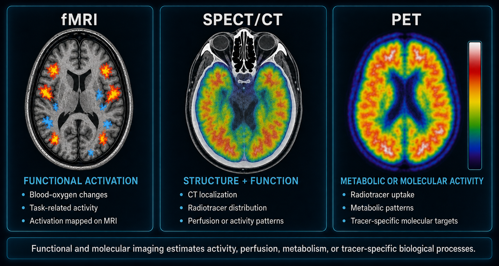
Structural imaging primarily asks, “What does the brain look like?” Functional and molecular imaging asks different questions: What is the brain doing? How is blood moving through it? How is it using oxygen or glucose? What molecular or chemical processes are occurring?
These technologies do not literally display thoughts, memories, emotions, intelligence, or pain. They measure biological signals associated with activity and use those measurements to create images or maps.
Functional Magnetic Resonance Imaging: fMRI
Functional MRI uses MRI technology to measure changes in blood oxygenation associated with brain activity. This is commonly called the blood-oxygen-level-dependent, or BOLD, signal.
When an area of the brain becomes more active, its oxygen demands change. fMRI detects related changes in oxygenated and deoxygenated blood and uses them to estimate which areas are participating in a task.
During a task-based fMRI, a person may be asked to move a hand or foot, read or speak, look at pictures, listen to sounds, respond to sensory stimulation, or complete memory and thinking tasks.
Clinical fMRI is frequently used to map language, movement, and other essential functions before brain surgery. It is also widely used in research involving cognition, behavior, pain, psychiatric conditions, brain injury, and neurological disease.
fMRI measures an indirect hemodynamic response, but it does not record individual neurons firing. Results can be affected by movement, breathing, medication, alertness, task performance, anxiety, and cardiovascular factors. An unusual activation pattern does not automatically identify the cause of symptoms or prove that an injury occurred.
Resting-State fMRI
Resting-state fMRI examines spontaneous BOLD-signal patterns while a person rests rather than completing a specific task. Researchers examine whether different brain areas demonstrate synchronized activity within networks involved in attention, memory, executive functioning, movement, sensory processing, emotional regulation, and self-referential thinking.
Resting-state fMRI is being studied in concussion and TBI because trauma may affect communication among brain regions even when structural imaging appears normal. Studies have reported both increased and decreased connectivity following TBI. These differences remain difficult to interpret consistently at the individual level (Lunkova et al., 2025).
Positron Emission Tomography: PET
Positron emission tomography is a nuclear-medicine procedure that uses a small amount of radioactive material called a radiotracer.
Different radiotracers are designed to participate in or attach to different biological processes. A specialized scanner detects energy released by the tracer and produces images showing where it accumulated. The commonly used FDG tracer is similar to glucose and can help estimate how actively different tissues are using glucose.
Brain PET may be used in selected evaluations involving dementia, epilepsy, brain tumors, abnormal metabolic patterns, neurodegenerative conditions, and neurological research.
PET provides molecular or metabolic information but generally has less anatomical detail than CT or MRI. PET is therefore commonly combined with CT—and sometimes MRI—so biological activity can be matched to anatomical structures (ACR & RSNA, 2025).
Single-Photon Emission Computed Tomography: SPECT
Single-photon emission computed tomography is another nuclear-medicine technique. It uses a radiotracer and gamma camera to create images showing tracer distribution. In brain imaging, SPECT frequently provides information about regional cerebral blood flow or perfusion.
Brain SPECT may be used in selected cases involving seizure localization, certain dementias, cerebrovascular disease, movement disorders, and other specialized neurological questions. SPECT is different from PET: the technologies use different tracers, detection systems, and protocols.
SPECT/CT
SPECT/CT combines functional information from SPECT with structural information from CT. The SPECT portion shows radiotracer distribution, while CT helps determine the anatomical location of that activity.
Some private clinics promote brain SPECT as a way to diagnose concussion, TBI, PTSD, or psychiatric conditions from blood-flow patterns. However, altered perfusion is not unique to traumatic brain injury. Similar patterns may be associated with medication, sleep problems, vascular conditions, psychiatric disorders, neurological disease, pain, and other factors.
SPECT may provide useful information for carefully selected clinical questions, but a SPECT image alone cannot prove that trauma caused a finding, determine when it developed, or establish that a normal scan excludes brain injury.
See our Spect CT page for more information.
PET/CT and PET/MRI
Most contemporary PET examinations are combined with CT. PET supplies molecular or metabolic information, while CT supplies anatomical information.
PET/MRI combines PET with MRI instead. This can provide detailed soft-tissue imaging alongside molecular measurements while reducing the CT-related portion of radiation exposure. However, the PET radiotracer still involves radiation. PET/MRI is not available everywhere and is generally used for selected neurological, oncological, or research purposes.
See our PET, CT, and MRI page for more information.
3. Electrical and Magnetic Activity

EEG and MEG are often grouped with functional imaging, but they are better described as neurophysiological recording and brain-mapping technologies. Unlike CT, MRI, PET, and SPECT, these tests focus on signals produced by neuronal activity.
One of their greatest strengths is temporal resolution: they can measure changes occurring within milliseconds, much faster than blood flow or metabolic imaging.
Electroencephalography: EEG
Electroencephalography records electrical activity using electrodes placed on the scalp. The electrodes do not send electricity into the brain during a standard diagnostic EEG. They detect electrical signals generated by neuronal activity and transmit them to recording equipment.
EEG is commonly used to evaluate epilepsy and seizures, episodes of altered awareness, unexplained loss of consciousness, encephalopathy, sleep disorders, certain causes of confusion, and brain activity in critically ill patients.
An EEG may be performed as a short routine study, sleep-deprived EEG, ambulatory recording, prolonged hospital monitoring, or video EEG, combining brain-wave recording with synchronized video.
EEG provides excellent information about when abnormal electrical activity occurs. Determining the exact source – especially when activity originates deep within the brain…can be more difficult. A routine EEG captures only the recording period, so intermittent abnormal activity may not occur during that window. A normal EEG does not rule out epilepsy, brain injury, cognitive impairment, persistent post-concussion symptoms, or every other neurological condition.
Quantitative EEG: qEEG
Quantitative EEG uses mathematical and statistical methods to analyze EEG recordings. It may compare features such as frequency, power, timing, or connectivity with a reference database.
qEEG is used in some clinical and research settings, but interpretation depends on recording quality, artifact removal, the normative database, medication, alertness, age, sleep, and technical settings. A colorful qEEG “brain map” is not a literal picture of damaged tissue. It is a visual representation of analyzed electrical measurements.
Magnetoencephalography: MEG
Magnetoencephalography measures the fragile magnetic fields produced by electrical activity in the brain. During conventional MEG, a person sits or lies with the head inside a helmet-shaped device containing sensitive sensors. These sensors passively detect magnetic fields; they do not produce radiation or inject energy into the brain.
MEG may be used to locate seizure activity; map language, movement, and sensory functions; assist with brain-surgery planning; study brain networks; and investigate neurological and psychiatric conditions.
EEG and MEG arise from related neuronal activity, but detect different physical signals. EEG measures electrical potentials at the scalp, while MEG detects associated magnetic fields (National Institute of Mental Health [NIMH], n.d.). MEG may offer improved source localization under favorable conditions, but it is expensive, technically complex, and available only at a limited number of specialized centers.
4. Experimental and Developing Imaging Technologies
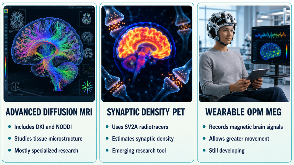
Brain-imaging research is moving beyond traditional pictures of anatomy. Scientists around the world are developing ways to examine white-matter complexity, brain connectivity, blood flow, tissue chemistry, metabolism, neuroinflammation, synaptic density, cellular ionic balance, tissue stiffness, molecular targets, and brain function during natural movement.
Some technologies in this section are already used clinically for selected conditions. What remains experimental may be their application to concussion, TBI, persistent symptoms, or another specific diagnosis.
“Experimental” does not mean imaginary, unreliable, or useless. It means researchers are still determining what an abnormal result means, whether it is specific to one condition, how results differ across equipment and institutions, what normal comparison values should be, and whether findings reliably change treatment or predict recovery.
Advanced Diffusion Imaging
DTI is only one method of examining diffusion. Newer approaches attempt to model the brain’s complicated tissue environment more accurately.
Diffusion Kurtosis Imaging: DKI
DKI evaluates how water movement differs from the simplified diffusion pattern assumed by conventional DTI. Researchers are investigating whether it can provide additional information about tissue complexity and microstructural disruption.
NODDI
Neurite orientation dispersion and density imaging uses mathematical modeling to estimate aspects of neurite density and organization. Neurites include portions of axons and dendrites. NODDI does not display individual nerve fibers or cells.
Free-Water Imaging
Free-water imaging attempts to separate extracellular water from water associated more closely with brain tissue. Researchers are investigating whether it may help characterize inflammation, swelling, degeneration, and other microstructural changes.
HARDI and Advanced Tractography
High-angular-resolution diffusion imaging, diffusion spectrum imaging, and constrained spherical deconvolution collect or analyze diffusion information in ways intended to represent complex and crossing fibers more accurately than basic DTI.
A 2026 study using DTI, free-water-corrected DTI, DKI, and NODDI found promising white-matter differences in people with subacute mild TBI. Larger studies and standardized clinical thresholds remain necessary before these methods can function as stand-alone diagnostic tests (Bergamino et al., 2026).
Susceptibility-Weighted Imaging and Quantitative Susceptibility Mapping
Susceptibility-weighted imaging, or SWI, is an MRI technique particularly sensitive to substances that alter the local magnetic field, including blood products and iron. It may reveal traumatic microbleeds difficult to see on CT or some routine MRI sequences. Microbleeds may provide evidence of traumatic vascular injury, but are not the same as direct proof of axonal shearing. Their absence does not rule out TBI.
Quantitative susceptibility mapping, or QSM, attempts to estimate magnetic-susceptibility differences within tissue. Researchers are investigating it for iron accumulation, small hemorrhages, myelin, venous structures, neurodegeneration, multiple sclerosis, and traumatic injury.
Arterial Spin Labeling: ASL
ASL is a noninvasive MRI technique that uses water in the person’s own arterial blood as a tracer. It can estimate cerebral blood flow without injecting a radioactive substance.
Researchers are studying ASL as a potential way to identify altered perfusion after concussion or TBI. Measurements may be affected by age, medication, sleep, caffeine, blood pressure, cardiovascular health, psychological conditions, and time since injury. Findings have not yet been consistent enough to make ASL an independent mild-TBI diagnostic test (Lunkova et al., 2025).
Magnetic Resonance Spectroscopy: MRS
MRS uses MRI technology to estimate concentrations of certain chemicals or metabolites in selected brain areas. Depending on the protocol, it may examine substances associated with neuronal integrity, energy metabolism, cell-membrane turnover, inflammation, glutamate and glutamine activity, and lactate production.
One commonly studied metabolite is N-acetylaspartate, or NAA. Decreased NAA may be associated with impaired neuronal or axonal integrity, but is not specific to traumatic injury. MRS may reveal biochemical differences in tissue that looks structurally normal, but does not directly display injured neurons or synapses.
A systematic review and meta-analysis found that TBI can affect brain metabolites, with NAA emerging as one of the more consistent findings. Results were still influenced by injury severity, brain region, timing, age, and imaging methods (Joyce et al., 2022).
Synaptic-Density PET: SV2A PET
One developing PET method targets synaptic vesicle glycoprotein 2A, or SV2A, a protein associated with presynaptic nerve terminals. Specialized radiotracers such as ¹¹C-UCB-J and ¹⁸F-SynVesT-1 bind to SV2A. Researchers use the PET signal as an estimate of synaptic density.
SV2A PET has been studied in epilepsy, Alzheimer’s disease, multiple sclerosis, depression, PTSD, neurodegeneration, and experimental traumatic-injury models. A foundational human study demonstrated that ¹¹C-UCB-J PET could estimate synaptic density in the living human brain (Finnema et al., 2016).
This may be the technology people mean when discussing imaging at the “synaptic level.” However, it estimates a synapse-associated protein rather than producing a microscopic picture of each synapse. It is not a routine clinical test for confirming an individual TBI after normal CT or MRI findings.
Neuroinflammation and Other Molecular PET Tracers
Researchers are developing PET tracers intended to identify processes related to neuroinflammation. Some target the translocator protein, or TSPO, which may increase in association with activated immune cells in the nervous system. TSPO is not specific to TBI; genetic differences affect binding, and an abnormal signal may not identify the precise cell type or cause.
Other tracers are being developed to examine amyloid, tau, myelin, dopamine systems, neurotransmitter receptors, cellular proliferation, and abnormal protein accumulation.
Ultra-High-Field MRI
Most clinical MRI scanners operate at 1.5 or 3 Tesla. Ultra-high-field scanners operate at 7 Tesla or higher. Seven-Tesla MRI can provide increased signal, improved contrast, and extremely detailed images of small vessels, cortical structures, microbleeds, epileptic lesions, multiple-sclerosis plaques, tumors, and pituitary abnormalities.
Seven-Tesla MRI has entered clinical use for selected indications, but access remains limited. Challenges include cost, specialized training, artifacts, implant restrictions, and uncertainty about whether greater detail improves outcomes for every condition. A European systematic review found promising neurological applications but noted that stronger evidence remains necessary for many indications (Radojewski et al., 2025).
Portable and Ultra-Low-Field MRI
Portable and ultra-low-field MRI systems use weaker magnetic fields and require less infrastructure than traditional MRI. Potential benefits include bedside imaging, intensive-care use, lower installation costs, and increased access in rural or underserved regions.
Tradeoffs may include lower resolution, weaker signal, longer scanning times, and fewer specialized sequences. In a 2025 European pilot study, portable ultra-low-field MRI showed promise in acute stroke care, although some very small lesions were missed (von Danwitz et al., 2025).
Wearable OPM-MEG
Conventional MEG uses large fixed systems containing extremely cold sensors. Optically pumped magnetometer MEG uses smaller quantum sensors positioned closer to the scalp.
Potential advantages include wearable adjustable helmets, greater comfort for children, recording while a person moves, and studying activity during more natural tasks. Researchers in the United Kingdom are developing mobile OPM-MEG to study acute neurological effects of blast exposure. Future applications may extend to military injuries, sports concussions, epilepsy, and dementia (UK Ministry of Defence, 2025).
Functional Near-Infrared Spectroscopy: fNIRS
fNIRS uses light transmitted through the scalp to estimate changes in oxygenated and deoxygenated blood near the brain’s surface. Equipment can be portable, allowing researchers to study people while walking, speaking, completing cognitive tasks, or participating in rehabilitation.
Limitations include restricted access to deeper brain structures, interference from hair, motion artifacts, scalp blood flow, skull thickness, and systemic cardiovascular effects. fNIRS is being studied in rehabilitation, concussion, developmental neuroscience, cognition, and bedside monitoring.
Functional Ultrasound: fUS
Functional ultrasound uses ultrafast ultrasound measurements to detect changes in cerebral blood flow or blood volume associated with neural activity. The adult skull is a major obstacle because bone blocks and distorts ultrasound.
Researchers in the Netherlands demonstrated mobile functional-ultrasound monitoring during walking in a person with an approved sonolucent skull implant. This was an important research milestone, but not a generally available noninvasive scan for the public (Soloukey et al., 2025).
Sodium MRI and CEST
Conventional MRI primarily detects hydrogen signals. Sodium MRI examines sodium distribution, which relates to cell-membrane integrity, ion-pump function, cellular energy, and tissue viability. It is being investigated in epilepsy, tumors, multiple sclerosis, stroke, neurodegeneration, and TBI. Challenges include low signal, long acquisition time, limited availability, and difficulty separating sodium inside cells from sodium outside cells (Egidi et al., 2025).
Chemical exchange saturation transfer, or CEST, uses interactions between water and other molecules to create contrast related to tissue chemistry. Researchers are investigating whether it can provide information about acidity, proteins, metabolism, tumors, and cellular changes. CEST is technically demanding and is not a routine TBI test.
Magnetic Resonance Elastography: MRE
MRE combines mechanical vibrations with MRI to estimate the stiffness and viscoelastic properties of brain tissue. Researchers are examining potential applications in aging, tumors, hydrocephalus, multiple sclerosis, neurodegeneration, and traumatic injury. MRE measures mechanical properties; it does not directly measure pain, cognition, or emotional distress (Jegathees et al., 2025).
Photon-Counting and Spectral CT
Photon-counting CT detects individual X-ray photons and records information about their energy. Potential advantages include higher spatial resolution, improved material differentiation, reduced noise, better imaging around bone or metal, detailed vessel imaging, and lower radiation exposure for some protocols.
Photon-counting CT is entering clinical practice, but researchers are still defining its neurological advantages. It remains a structural X-ray technology and should not be expected to display synaptic injury.
Artificial Intelligence and Multimodal Analysis
Artificial intelligence is not an imaging modality by itself. It is increasingly used to process CT, MRI, PET, SPECT, EEG, and MEG data through automated lesion detection, image reconstruction, motion correction, brain-volume measurement, longitudinal comparison, pattern recognition, and multimodal data integration.
Concerns include biased training data, false positives, limited transparency, privacy, lack of external validation, and poor performance on populations or scanners unlike those used during development. An AI-generated or synthetic image must never be represented as a direct measurement of anatomy or biological activity that was not actually scanned.
Earlier-Stage Technologies
Additional technologies remain largely in engineering, animal, or early human research:
- Photoacoustic imaging, combining light and ultrasound
- Magnetic particle imaging, detecting magnetic nanoparticle tracers
- Electrical impedance tomography, estimating differences in electrical conductivity
- Microwave brain imaging, studying tissue through electromagnetic waves
- Advanced quantum sensors for more sensitive magnetic measurements
- Multimodal fusion combining several imaging and physiological sources
- AI-generated cross-modality imaging that estimates one image type from another
These approaches may eventually contribute to care, but their appearance in research or advertising is still in the developmental process and not yet readily available.
5. Miscellaneous: Biofeedback, Neurofeedback, and Brain X-rays
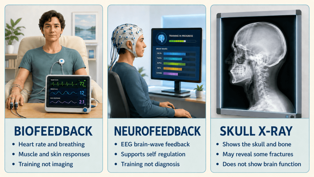
Some technologies related to the brain do not fit neatly into structural imaging, functional or molecular imaging, or electrical and magnetic diagnostic recording.
Biofeedback
Biofeedback is not a brain-imaging test. It is a training approach that uses sensors to measure physiological activity and return that information to the person in real time.
Biofeedback may monitor heart rate, heart-rate variability, breathing, muscle tension, skin temperature, skin conductance, or brain-wave activity. Information may be displayed through graphs, sounds, lights, animations, or computer exercises. The person uses the feedback to practice changing a physiological response.
The goal is not to create a picture of an injury. The goal is to improve awareness and, in some cases, develop greater voluntary regulation of bodily responses.
Neurofeedback
Neurofeedback is a specific form of biofeedback involving brain-wave measurements, usually obtained through EEG electrodes on the scalp. Software analyzes selected EEG features and provides visual or auditory feedback.
Neurofeedback does not read thoughts, produce a structural brain image, directly show damaged tissue, confirm that a TBI occurred, replace a clinical EEG, or establish a diagnosis by itself.
A qEEG may sometimes help develop a neurofeedback protocol, but a colorful brain map should not be mistaken for a CT, MRI, or direct picture of an injured brain. Evidence varies by condition, protocol, provider qualifications, and outcome. Patients should ask about practitioner credentials, supporting evidence, treatment goals, and how progress will be evaluated (Mayo Clinic, 2025).
Brain or Skull X-Rays
The phrase “brain X-ray” is misleading because ordinary X-rays do not provide useful images of brain tissue. Traditional skull X-rays primarily show dense structures such as bone. They may display certain fractures, bone abnormalities, foreign objects, dental or facial structures, or some calcifications.
Skull X-rays provide far less information about head trauma than CT. CT creates cross-sectional images and can show the skull, bleeding, swelling, and other internal abnormalities much more effectively. For this reason, ordinary skull X-rays are generally not the preferred imaging study for evaluating traumatic brain injury.
An X-ray cannot rule out concussion, intracranial bleeding, brain swelling, traumatic axonal injury, functional disruption, or cognitive impairment. When someone says they received a “brain X-ray,” it is worth asking whether the examination was actually a skull X-ray, head CT, or another study.
The Holistic Approach and An Important Lesson
Every single test provides a specific picture of brain or skull health.
These are the 3 most commonly used:
- CT is fast and especially useful for acute bleeding and fractures (why this is often the one chosen during emergency room visits)
- MRI provides detailed structural information (sometimes used in emergency room visits, but not as often as CT)
- Ordinary X-rays primarily show bone (skull) and not the brain itself.
These are usually referred to a specialist by an emergency room physician or a primary care physician (PCP):
- DTI evaluates aspects of white-matter diffusion.
- fMRI estimates activity-related blood-oxygenation changes.
- PET evaluates metabolism or other molecular targets.
- SPECT evaluates radiotracer distribution and regional perfusion.
- EEG records electrical activity.
- MEG records associated magnetic fields.
- Emerging technologies examine chemistry, connectivity, inflammation, synaptic density, ionic balance, and tissue mechanics.
- Biofeedback and neurofeedback use physiological information for training rather than diagnostic imaging.
A normal CT or MRI scan does not automatically invalidate a person’s symptoms. At the same time, an abnormal or colorful image does not automatically prove what caused the finding.
Brain injury should be evaluated and treated through a holistic (whole-person, individualized) approach…not by treating the scan alone. Two people exposed to similar forces may have completely unique symptoms, medical histories, vulnerabilities, and recovery paths. Loss of consciousness is not required for a concussion, and a person may sustain clinically meaningful disruption in their lives even when there is no skull fracture, visible bleeding, or other acute abnormality on routine CT or MRI. Cellular, axonal, metabolic, or synaptic-level dysfunction may be too subtle for those structural studies to display directly.
A thorough history is absolutely imperative as part of the individual documentation. This approach should consider the mechanism of injury or circumstances and timing of the current injury; symptoms that appeared immediately, later, or are persisting; any loss of consciousness and length of time loss lasted, confusion, memory gaps, or seizure; and the person’s lifetime history of diagnosed or suspected concussions and other head impacts. Recent injuries are especially important because recovery may be slower in people who have experienced a previous concussion or TBI, and another injury during recovery may complicate symptoms and rehabilitation (Centers for Disease Control and Prevention [CDC], 2025a, 2025b).
Sex and gender may also influence how brain injury is experienced, recognized, treated, and supported. Research…particularly in mild TBI and sports concussion…has found that female survivors often report more symptoms, more severe symptoms, and in some areas, a longer recovery than male survivors. A recent systematic review reported differences involving pain, cognition, balance and gait, vestibular and visual symptoms, return to activity, cerebral blood flow, and white-matter findings (Arachchi et al., 2026). Biological factors such as hormones and neurochemistry may contribute, while gender-related differences in injury exposure, symptom reporting, medical recognition, caregiving demands, social support, and access to appropriate treatment may also affect outcomes.
PINK Concussions highlights that women and girls can face additional doubt, isolation, or pressure when their symptoms or recovery do not match expectations formed around male experiences (PINK Concussions, n.d.). These are group-level patterns, not rules for an individual: not every female patient will have a prolonged recovery, and male and gender-diverse patients can also experience severe or persistent effects. While sex and gender should still be considered, every person’s individual symptoms and experiences must be heard and evaluated.
The assessment should also consider the person’s health before the injury and any conditions that developed, changed, or worsened afterward. Sleep disorders, chronic pain, migraine, medication effects, mental-health conditions, cervical injuries, vestibular or vision problems, and endocrine, inflammatory, or autoimmune conditions may overlap with, mimic, intensify, or be generated by post-traumatic symptoms. These conditions should neither be dismissed as “just the brain injury” nor used automatically as “preexisting” to deny that an injury occurred. Instead, clinicians should investigate how the factors may be interacting in that individual.
Traumatic brain injury can also disrupt the hypothalamic-pituitary system and lead to post-traumatic hormone deficiencies in some people. Symptoms can overlap substantially with common TBI complaints and may include persistent fatigue, cognitive difficulty, mood changes, altered weight, menstrual or sexual function changes, thirst or urination changes, poor rehabilitation progress, or the appearance of inconsistent effort. Some patients…particularly those with moderate or severe TBI, or people with milder injury who have symptoms suggestive of pituitary dysfunction may require targeted endocrine evaluation rather than assumptions based on imaging alone (Tan et al., 2017).
The goal is not simply to decide whether a picture looks “normal.” The goal is to understand what happened to this particular person, identify treatable contributors, and develop a coordinated plan that addresses the whole person.
Understanding the “alphabet soup” gives us a starting point. It helps us ask informed questions, recognize the limitations of individual tests, communicate more effectively with healthcare professionals, and become active and informed participants in our care, or in the care of those we care about or are advocating for.
Self Advocacy: 12 Suggested Questions That May Be Helpful To Ask
The goal of informed advocacy is not to demand the newest or most expensive test. It is to understand the clinical question and whether the proposed test is appropriate for answering it.
- What specific question is this test intended to answer?
- Is it examining anatomy, blood flow, metabolism, electrical activity, magnetic signals, or another biological process?
- Why was this test selected instead of another option?
- What findings can it reliably detect?
- What can it not rule out?
- Could medication, sleep, movement, another diagnosis, or another health condition affect the result?
- Does it have an accepted clinical role for my condition, or is this use experimental?
- Who will interpret the study, and what specialized training does that person have?
- Will the result change my diagnosis, treatment, rehabilitation, or follow-up plan?
- If the result is normal but my symptoms continue, what happens next?
- If it is abnormal, is the finding specific to my injury or condition?
- Will insurance cover the test?
References used in compilation of this post:
American College of Radiology, & Radiological Society of North America. (2025). PET/CT—positron emission tomography/computed tomography. RadiologyInfo.org. https://www.radiologyinfo.org/en/info/pet
American College of Radiology, & Radiological Society of North America. (2026a). Head CT. RadiologyInfo.org. https://www.radiologyinfo.org/en/info/headct
American College of Radiology, & Radiological Society of North America. (2026b). Brain MRI. RadiologyInfo.org. https://www.radiologyinfo.org/en/info/mri-brain
Asken, B. M., DeKosky, S. T., Clugston, J. R., Jaffee, M. S., & Bauer, R. M. (2018). Diffusion tensor imaging findings in adult civilian, military, and sport-related mild traumatic brain injury: A systematic critical review. Brain Imaging and Behavior, 12, 585–612. https://pubmed.ncbi.nlm.nih.gov/28337734/
Bergamino, M., Ott, F. W., McElvogue, E. J., Jha, R. M., Moreno, S., & Stokes, A. M. (2026). Multimodal diffusion MRI biomarkers of white matter alterations and clinical impairment in mild traumatic brain injury. https://pubmed.ncbi.nlm.nih.gov/42292335/
Brain Injury Association of America. (n.d.). About brain injury. https://biausa.org/brain-injury/about-brain-injury
Egidi, I., Guidi, M., & Giove, F. (2025). Compartmentalization of sodium in the human brain: A mini-review of ²³Na-MRI methods. Frontiers in Physics, 13, Article 1487822. https://doi.org/10.3389/fphy.2025.1487822
Finnema, S. J., Nabulsi, N. B., Eid, T., Detyniecki, K., Lin, S.-F., Chen, M.-K., Dhaher, R., Matuskey, D., Baum, E., Holden, D., Spencer, D. D., Mercier, J., Hannestad, J., Huang, Y., & Carson, R. E. (2016). Imaging synaptic density in the living human brain. Science Translational Medicine, 8(348), 348ra96. https://doi.org/10.1126/scitranslmed.aaf6667
HOPE TBI. (n.d.). HOPE TBI. https://hopetbi.com/
Jegathees, T., Jugé, L., Hau, E., Bilston, L. E., & O’Neill, G. M. (2025). The glioblastoma biomechanical landscape: A systematic review of magnetic resonance elastography of brain tumors and healthy brain. APL Bioengineering, 9(3), 031503. https://pubmed.ncbi.nlm.nih.gov/40949715/
Joyce, J. M., La, P. L., Walker, R., & Harris, A. D. (2022). Magnetic resonance spectroscopy of traumatic brain injury and subconcussive hits: A systematic review and meta-analysis. Journal of Neurotrauma. https://doi.org/10.1089/neu.2022.0125
Lunkova, E., Chen, J.-K., McCabe, S., Saluja, R. S., & Ptito, A. (2025). Multimodal approach in the identification of biomarkers of mild traumatic brain injury: Resting-state fMRI, ASL, and SWI. Clinical Neuroimaging, 2(1), Article e70026. https://doi.org/10.1002/neo2.70026
Mayo Clinic. (2025). Biofeedback. https://www.mayoclinic.org/tests-procedures/biofeedback/about/pac-20384664
PINK Concussions. (n.d.). Female brain injury. https://www.pinkconcussions.com/brain-injury
Disclaimer
HOPE TBI is a public educational and advocacy website, not a legal entity, or an academic journal. Sources may be presented through hyperlinks or simplified references rather than strict academic citation formatting. Readers should consult and cite the original sources when using this material for academic, legal, clinical, or professional purposes. Information reflects sources available as of the publication or latest update date. Medical knowledge and recommendations may change. Sources are provided for verification, and corrections are welcome. If you identify an error, outdated link, inaccurate quotation, or needed correction, please CONTACT US so we can review and update the material.
 HOPE TBI